Zebrafish Obesogenic Test, label‑free
ViZOT quantifies anal fin ray and ventral subcutaneous adipocytes directly on the brightfield image acquired at each ZOT session, in one step: no crop, no background subtraction, no threshold to tune.
Label-free Adiposity & ZOT Methodology
Obesogens promote fat storage, and some of them induce a thrifty phenotype in which adiposity is preserved despite fasting. The ZOT identifies such compounds in vivo by measuring, in the same larva, the area of subcutaneous adipocytes before and after a 24-h exposure in a fasting state. Staining every larva twice with Nile Red adds a reagent that may interact with the tested compounds and limits repeated measurements on the same animal.
Whole-Organism ZOT Assay
Larvae of 8.5 to 9.5 mm standard length are imaged before and after a 24-h exposure in a fasting state, and each larva is its own reference for the adiposity variation.
Brightfield Acquisition
Adipocyte lipid droplets appear as refractile spheres at the 4× magnification of the ZOT. The brightfield image is already the first channel of every ZOT acquisition.
One-Step U-Net
A single U-Net with a ResNet-34 encoder segments AFRSAT and VSAT adipocytes on the whole image. ViZOT-BF reads the brightfield image, ViZOT-NR the Nile Red fluorescence image.
ZOTAS-Compatible Readout
Adipocyte areas in pixels and mm², area variation normalized to the controls and statistics versus the control, in the folder layout and Excel tables of ZOTAS.
Same conclusions as ZOTAS
The larvae of the published screen were reanalyzed with ViZOT-NR and ViZOT-BF and compared, larva by larva, with the published ZOTAS measurements.
Compounds, 28 Experiments
486 larvae for AFRSAT and 500 for VSAT, analyzed with the three methods on identical sets of animals.
Obesogens Identified
Amiodarone, rosiglitazone, triclosan and dibutyl phthalate induced a thrifty phenotype with the three methods (p < 0.0001); the eight other compounds showed no significant difference.
Unseen Compounds Classified
Amiodarone (obesogenic) and DEHP (non-obesogenic), withheld from training and validation, were correctly classified by both models.
Excluded Larvae Recovered
All the larvae with images that were left out of the published analyses (ZOTAS threshold failure, manual exclusion) were quantified, in one step and without operator intervention.
Associated Scientific Publications
The ZOT bioassay, its optimized protocol and the ZOTAS software, and the 2026 screen whose larvae were reanalyzed with ViZOT, which identified the same obesogens as ZOTAS.
Area variations measured with ViZOT were correlated with those of ZOTAS (Spearman r = 0.98 and 0.96 for ViZOT-NR, 0.88 and 0.86 for ViZOT-BF, AFRSAT and VSAT), areas on single images even more closely (r ≥ 0.98, slopes 0.95 to 1.03), and the dispersion of the area variation among control larvae was similar with the three methods.
How ViZOT Works
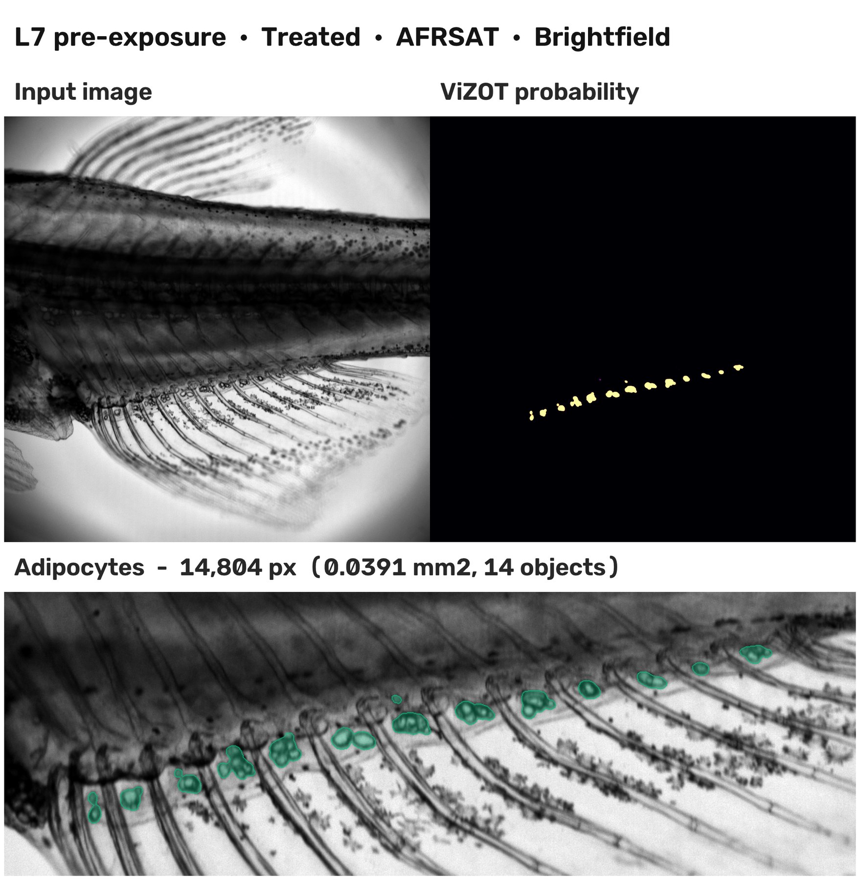
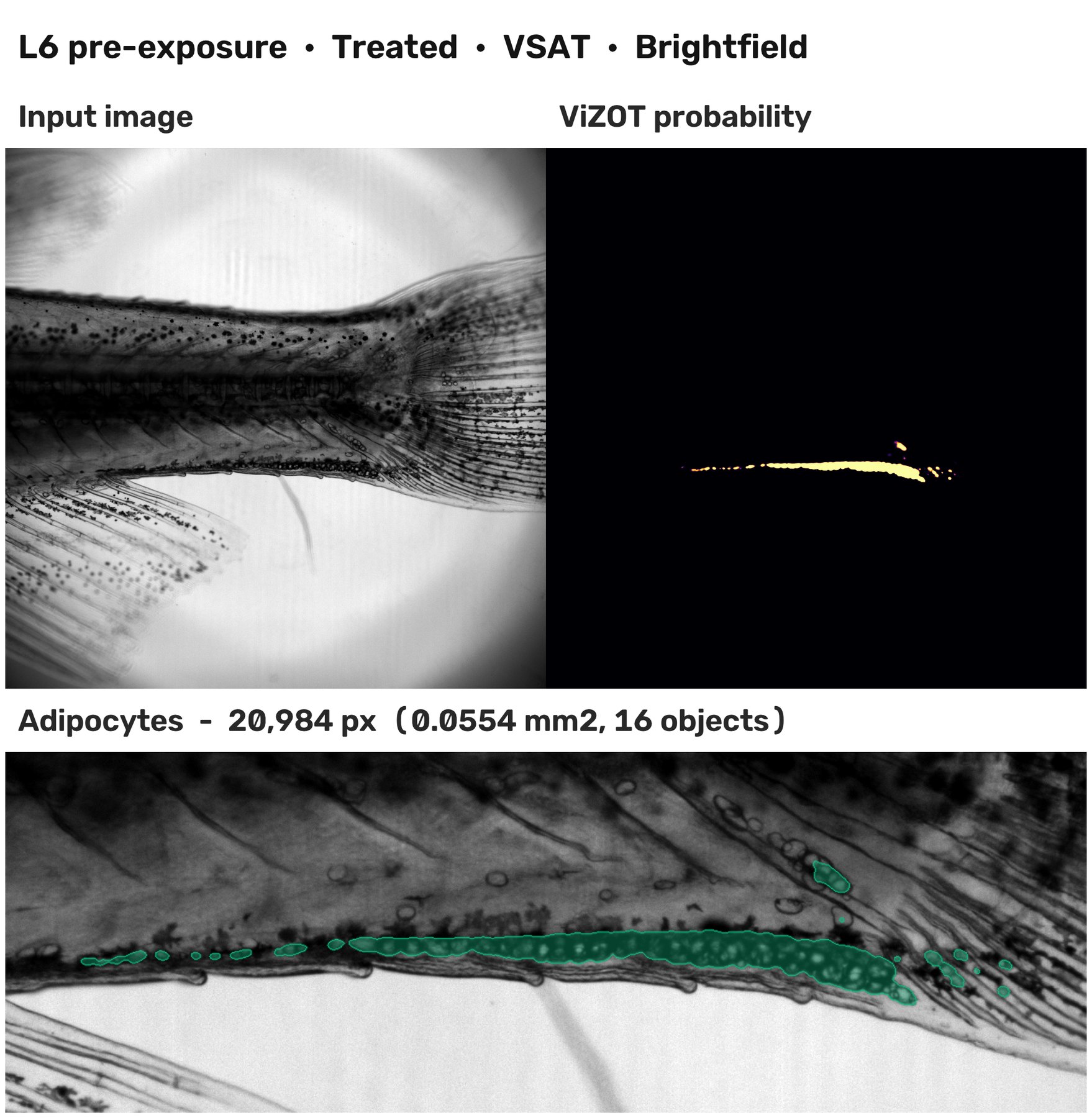
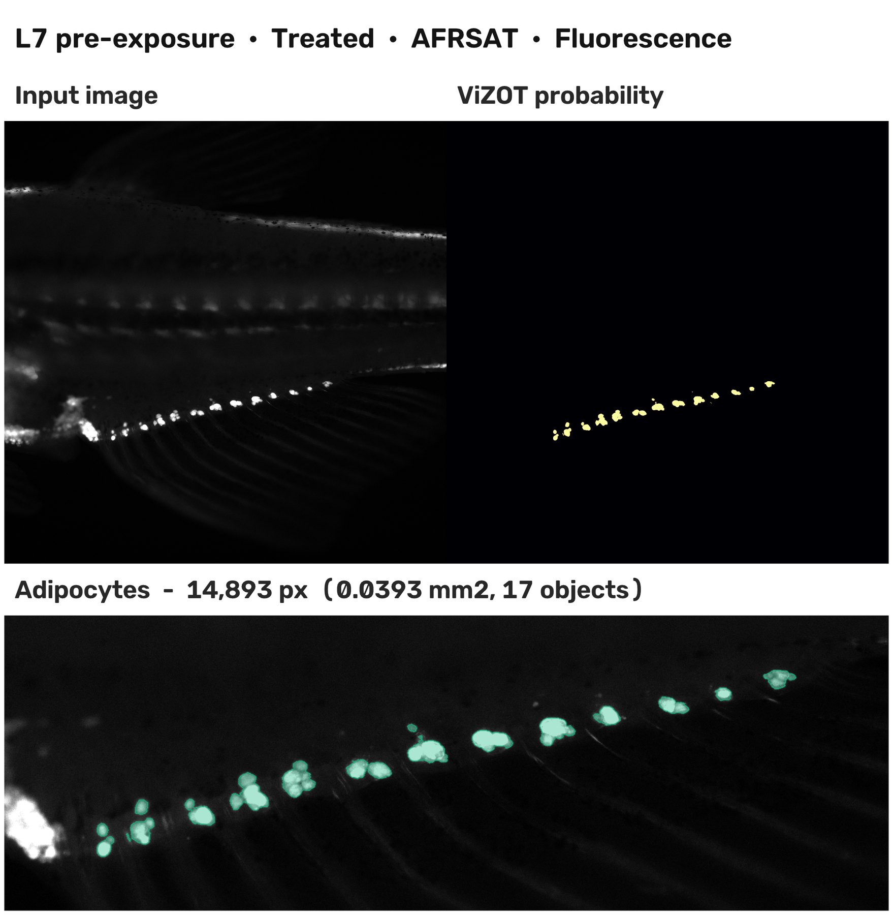
Processing plots written by the desktop application for one larva: input image, ViZOT probability map and the adipocytes it measures.
Brightfield AFRSAT
ViZOT-BF finds the anal fin ray adipocytes on the whole brightfield frame, without crop or threshold selection.
Brightfield VSAT
The same network measures the ventral adipocytes: one image per depot, areas in pixels and mm².
Nile Red
ViZOT-NR reads the Nile Red fluorescence image of the same acquisition, for experiments analyzed with both channels.

Download ViZOT
The desktop application runs the ViZOT-BF and ViZOT-NR networks on whole ZOT experiments, offline: one installer for every Windows PC, using the NVIDIA GPU when it is faster than the processor.
Downloads are currently restricted to authorized accounts. After publication, ViZOT will be available to registered users once their registration has been validated.
ViZOT was trained on images from one set-up (Nikon Eclipse Ni, 4× Plan Apo, ORCA-Flash4.0); other microscopes and cameras remain to be evaluated.
Explore ViZOT-BF & ViZOT-NR
The training and inference code and the weights of both models will be released with the article.
For help adapting ViZOT to your own microscope, contact theo.merce@danio-tracker.com .
Credits & Institutional Partners